Database Open Access
PhysioZoo - mammalian NSR databases
Published: Aug. 27, 2019. Version: 1.0.0
When using this resource, please cite:
Shemla, O., & Behar, J. (2019). PhysioZoo - mammalian NSR databases (version 1.0.0). PhysioNet. RRID:SCR_007345. https://doi.org/10.13026/p63q-hq95
Please include the standard citation for PhysioNet:
(show more options)
Goldberger, A., Amaral, L., Glass, L., Hausdorff, J., Ivanov, P. C., Mark, R., ... & Stanley, H. E. (2000). PhysioBank, PhysioToolkit, and PhysioNet: Components of a new research resource for complex physiologic signals. Circulation [Online]. 101 (23), pp. e215–e220. RRID:SCR_007345.
Abstract
The PhysioZoo database contains electrocardiographic recordings (ECG) taken from multiple types of mammals (dogs, rabbits, mice). Each record is provided in 3 different formats: MATLAB (.mat), simple text (.txt), PhysioNet (.hea,.dat,.qrs). For each record the following data is available: the raw ECG signal, reference (human corrected) R-peak annotations and the signal quality annotations. All of the files formats are compatible with the PhysioZoo open source software available at physiozoo.com. When the recordings were taken, all the mammals were conscious, and no drugs were administered prior to the recordings.
Background
Over the past few decades, numerous studies have explored the variation of the time interval between heartbeats, also known as heart rate variability (HRV). Studies have shown that measures quantifying HRV together with heart rate (HR) can provide useful information on cardiovascular health [1-3].
With the recent advances in genome manipulation technologies, animals with mutations designed to overexpress or knock out genes implicated in human cardiovascular diseases have become an important focus of biological research [4]. HRV is a non-invasive tool that can be used to analyze the electrical heart activity of mutant animals and provide new insights into the pathophysiology and how these conditions may be diagnosed. HRV analysis has also been used to study the role of cardiac mediators and signaling pathways in heart rhythm [5,6] and the effect of pharmacological substances on the HRV [7,8]. Such experiments can only be performed using animal models.
Despite the clear motivation for integrating HRV analysis into animal studies, some drawbacks prevent researchers from doing so: (i) there are no standard publicly available R-peak detector algorithms adapted for use with mammalian electrophysiological data; (ii) the HRV measures and available software implementing them are based on human electrophysiological data analysis, and there is no standard method for adapting them to other mammals; and (iii) there is no standardized annotated database that can be used as a reference when developing new programs for HRV analysis or new HRV indices. These limitations have motivated the creation of the PhysioZoo platform for HRV analysis of mammalian electrophysiological data. This new platform includes a set of standardized databases of electrophysiological data from mammals classicaly used in research, R-peak detectors for accurately estimating the beat-to-beat intervals from electrophysiological recordings of different mammals, and software implementing the state of the art HRV measures, with parameters adapted to each species.
The PhysioZoo platform enables the standardization and reproducibility of HRV analysis for human, dog, rabbit, and mouse electrocardiographic (ECG) data through its open source code, freely available software, and open access ECG databases. PhysioZoo will support and enable new developments in mammalian HRV research, and new contributions of mammalian electrophysiological recordings.
Methods
Electrocardiographic data from dogs [9], rabbits [10,11], and mice [12] were obtained. All animal data used were obtained from published studies for which the respective animal protocols and experimental procedures were approved by the original research committee [9-13]. Dog data were recorded at 500 Hz, and body surface electrodes were placed on either side of the animal’s chest and secured with surgical tape. Rabbit data were recorded by means of subcutaneous ECG recording using the Ponemah platform (DSI, MN, United States) at 1 kHz. ECG raw data were exported to text files from the Ponemah software using the maximal precision (four decimal places). All rabbits were female and free moving in a cage. Mouse data were recorded using Telemetry sensors (ETA-F20 or HDX-11, Data Sciences International, Saint Paul, MN, United States) with a sampling rate of 1000 Hz. All the mammals were conscious, and no drugs were administered prior to the recording.
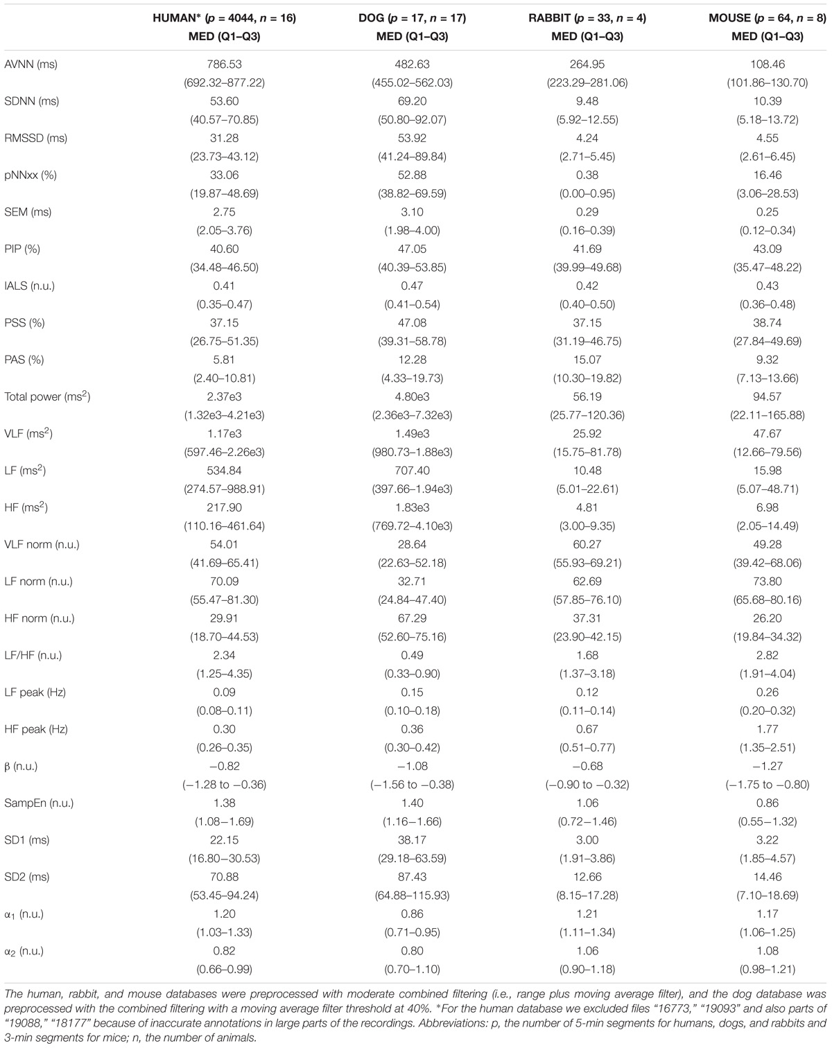
We performed peak detection to identify the R-peak locations [14] in the animal databases. Because no state of the art R-peak detector has ever been designed and evaluated for data from animals (whose beating rates differ from that of humans), we manually corrected the peak locations. For that purpose, a single trained annotator reviewed all the recordings and corrected the inaccurate annotations (false positive and false negative). In addition, segments were marked as bad quality when no R-peak could be visually identified by the annotator for at least three consecutive peaks. Annotations within these low quality segments were removed. Thus, for each record in our database, we obtained the reference (i.e., human corrected) R-peak annotations and the signal quality annotations. We used these reference annotations to evaluate the mammal-specific R-peak detector implemented in the PhysioZoo software as well as to provide standard ranges of the HRV measures, available in https://www.frontiersin.org/files/Articles/391349/fphys-09-01390-HTML/image_m/fphys-09-01390-t006.jpg
Data Description
Databases summary
A summary of the records in the database is given below:
|
Dog
|
Rabbit
|
Mouse
|
|
|
Number of records |
17 |
20 |
8 |
|
Number of mammals |
17 |
4 |
8 |
|
Average length (hr:min:sec) |
00:05:31 |
00:10:34 |
00:29:44 |
|
Min length (hr:min:sec) |
00:04:09 |
00:04:53 |
00:13:53 |
|
Max length (hr:min:sec) |
00:06:48 |
00:26:00 |
00:39:38 |
|
Total length (hr:min:sec) |
01:33:55 |
03:31:13 |
03:28:07 |
|
Total R-peak annotations |
10,871 |
50,452 |
109,865 |
|
Bad quality, Gross (%) |
2.7% |
1.0% |
0.4% |
The files in these databases are given in 3 types of formats:
- WFDB (Physionet's .hea,.dat,.qrs),
- text (.txt),
- MATLAB (.mat).
The .txt and .mat files structures are specific to PhysioZoo whereas the WFDB follows the standard structure of WFDB powered by Physionet (physionet.org) with some additional fields in the WFDB header files. If you are experienced with WFDB then we recommend you use this format.
There are 3 types of data files given for the records in these databases:
- ECG time series,
- The location of the peaks (e.g. R-peak from an ECG time series),
- Annotation on the signal quality.
For any of the three formats a file consists of a “header” and the “data”.
General structure of the Header
An extensive description of the header structure and examples are provide in the PhysioZoo documentation.
For each of the files the following information can be specified in the header:
- Mammal: the name of the mammal (e.g. ‘dog’, ‘mouse’, ‘rabbit’, ‘human’),
- Fs: the sampling frequency in Hertz,
- Integration_Level: ‘electrocardiogram’, ‘electrogram’ or ‘action potential’.
As part of the header, information characterizing each channel available can be entered:
- type: ‘peak’, ‘signal quality’, ‘time’, ‘interval’, ‘beating rate’ and ‘electrography’. See definitions of these in the next section,
- name: a name you want to give to the channel (of note this information is not used by the PZ Loader to load the data),
- unit: ‘millisecond’, ‘second’, ‘index’, ‘bpm’, ‘millivolt’, ‘microvolt’ and ‘volt’,
- enable: ‘yes’ or ‘no’. If you specify ‘no’ then this channel will be ignored when the file is loaded by the PZ Loader. Only specify ‘yes’ for the channels you want to use.
In the case all or some of this information is missing from the header then you will be prompted to enter it through the PZ Loader User Interface.
Channels type
There are two categories of Channels: ‘Annotations’ and ‘Time series’. Within these two categories the Channel can be one of the following type’s (specified in the header):
- Annotations
-
- ‘peak’: the location of the peaks (e.g. R-peak from an ECG time series). The peak locations can be specified in millisecond/second or index,
- ‘signal quality’: annotation on the signal quality. The signal quality annotations can be specified in millisecond/second or index.
- Time series
-
- ‘time’: a time vector giving the position of each sample of another time series. An entry of type ‘time’ needs to be associated with another time series and must be in units of seconds or milliseconds,
- ‘interval’: the time interval between consecutive beats (e.g. RR time series). The interval length can be specified in second/millisecond or index,
- ‘beating rate’: the reciprocal of the interval in units of beats per minute (e.g. heart rate),
- ‘electrography’: the amplitude of an electrography time series (e.g. ECG). The electrography amplitude is given in microvolt, millivolt or volt. Thus only physiological units are allowed.
Headers in the different formats
Text format (.txt)
The header in the text files is given at the beginning of the file, such as:
---
Mammal: dog
Fs: 500
Integration_level: electrocardiogram

Channels:

    - type:   electrography
      name:   Data
      unit:   mV
      enable: yes

---


0.060057
0.064323
0.068589
Matlab format (.mat)
Each .mat file needs to contain the following fields:
- 'Data': a numeric vector containing the data in the file,
- 'Mammal': a string, the name of the mammal ('dog'/'rabbit'/'mouse'),
- 'Fs': a numeric scalar, the measurement frequency (500/1000),
- 'Integration_level': a string, describing the level of integration ('electrocardiogram'),
- 'Channels': a Cell array. Each element of the Channel Cell will contain the following fields : ‘type’, ‘name’, ‘unit’ and ‘enable’.
WFDB (.hea)
WFDB files (annotation or data) will be accompanied by a header (.hea) file specifying the relevant information for reading an annotation or data file. Refer to the WFDB documentation for that. To the standard WFDB format of the header, you will need to add one comment line at the end of the header, starting with a ‘#’ then followed by the mammal type and the integration level. For instance:
Dog_01 1 500 134258
Dog_01.dat 16 51300.0027(-1159)/mV 0 0 -1487 25886 0 Data (electrography)
#Mammal:dog,Integration_level:electrocardiogram
Usage Notes
An extensive description of the files formats, structure and examples are provided in the PhysioZoo documentation. The PhysioZoo software is available at: https://physiozoo.com/
Acknowledgements
The work was supported by the Center for Absorption in Science, Ministry of Aliyah and Immigrant Absorption, State of Israel (JB), the National Natural Science Foundation of China/Israel Science Foundation Joint Research Program, no. 398/14 (YY), Ministry of Science and Technology, Israel (YY), 5R01HL110791-05 (GK), T32AG041688 (KM, PI John Sedivy) The funders had no role in study design, data collection and analysis, decision to publish, or preparation of the manuscript.
Conflicts of Interest
The authors have no conflicts of interest to declare.
References
- Yaniv, Y., Ahmet, I., Tsutsui, K., Behar, J., Moen, J. M., Okamoto, Y., . . . Lakatta, E. G. (2016). Deterioration of autonomic neuronal receptor signaling and mechanisms intrinsic to heart pacemaker cells contribute to age-associated alterations in heart rate variabilityin vivo. Aging Cell, 15(4), 716-724. doi:10.1111/acel.12483
- Yaniv, Y., & Maltsev, V. A. (2014). Numerical Modeling Calcium and CaMKII Effects in the SA Node. Frontiers in Pharmacology, 5. doi:10.3389/fphar.2014.00058
- Odening, K. E., Choi, B., Liu, G. X., Hartmann, K., Ziv, O., Chaves, L., . . . Koren, G. (2012). Estradiol promotes sudden cardiac death in transgenic long QT type 2 rabbits while progesterone is protective. Heart Rhythm, 9(5), 823-832. doi:10.1016/j.hrthm.2012.01.009
- Brunner, M., Peng, X., Liu, G. X., Ren, X., Ziv, O., Choi, B., . . . Koren, G. (2008). Mechanisms of cardiac arrhythmias and sudden death in transgenic rabbits with long QT syndrome. Journal of Clinical Investigation. doi:10.1172/jci33578
- Billman, G. E. (2013). The effect of heart rate on the heart rate variability response to autonomic interventions. Frontiers in Physiology, 4. doi:10.3389/fphys.2013.00222
- Gross, V., Tank, J., Obst, M., Jordan, J., Diedrich, A., Plehm, R., & Luft, F. C. (2004). Clonidine Improves Baroreflex Sensitivity In Mice Through Parasympathetic Activation. Journal of Hypertension, 22(Suppl. 2). doi:10.1097/00004872-200406002-00075
- Elghozi, J., Girard, A., & Laude, D. (2001). Effects of drugs on the autonomic control of short-term heart rate variability. Autonomic Neuroscience, 90(1-2), 116-121. doi:10.1016/s1566-0702(01)00276-4
- Witte, K., Engelhardt, S., Janssen, B. J., Lohse, M., & Lemmer, B. (2004). Circadian and Short-Term Regulation of Blood Pressure and Heart Rate in Transgenic Mice with Cardiac Overexpression of The β1-Adrenoceptor. Chronobiology International, 21(2), 205-216. doi:10.1081/cbi-120037801
- Uechi, M., Asai, K., Osaka, M., Smith, A., Sato, N., Wagner, T. E., . . . Vatner, S. F. (1998). Depressed Heart Rate Variability and Arterial Baroreflex in Conscious Transgenic Mice With Overexpression of Cardiac G sα. Circulation Research, 82(4), 416-423. doi:10.1161/01.res.82.4.416
- Thireau, J., Zhang, B. L., Poisson, D., & Babuty, D. (2007). Heart rate variability in mice: A theoretical and practical guide. Experimental Physiology, 93(1), 83-94. doi:10.1113/expphysiol.2007.040733
- Yaniv, Y., Tsutsui, K., & Lakatta, E. G. (2015). Potential effects of intrinsic heart pacemaker cell mechanisms on dysrhythmic cardiac action potential firing. Frontiers in Physiology, 6. doi:10.3389/fphys.2015.00047
- Pincus, S. M., & Goldberger, A. L. (1994). Physiological time-series analysis: What does regularity quantify? American Journal of Physiology-Heart and Circulatory Physiology, 266(4). doi:10.1152/ajpheart.1994.266.4.h1643
- Behar, J. A., Rosenberg, A. A., Shemla, O., Murphy, K. R., Koren, G., Billman, G. E., & Yaniv, Y. (2018). A Universal Scaling Relation for Defining Power Spectral Bands in Mammalian Heart Rate Variability Analysis. Frontiers in Physiology, 9. doi:10.3389/fphys.2018.01001
- Behar, J., Oster, J., & Clifford, G. D. (2014). Combining and benchmarking methods of foetal ECG extraction without maternal or scalp electrode data. Physiological Measurement, 35(8), 1569-1589. doi:10.1088/0967-3334/35/8/1569
Access
Access Policy:
Anyone can access the files, as long as they conform to the terms of the specified license.
License (for files):
Open Data Commons Attribution License v1.0
Discovery
DOI (version 1.0.0):
https://doi.org/10.13026/p63q-hq95
DOI (latest version):
https://doi.org/10.13026/p19w-dp86
Topics:
heart rate variabillity
electrophysiology
mammals
ecg
Project Website:
https://physiozoo.com/
Project Views
151
Current Version151
All VersionsCorresponding Author
Versions
Files
Total uncompressed size: 401.3 MB.
Access the files
- Download the ZIP file (117.3 MB)
-
Download the files using your terminal:
wget -r -N -c -np https://physionet.org/files/physiozoo/1.0.0/
-
Download the files using AWS command line tools:
aws s3 sync --no-sign-request s3://physionet-open/physiozoo/1.0.0/ DESTINATION
| Name | Size | Modified |
|---|---|---|
| Parent Directory | ||
| Data_Quality_Annotations_Mouse_03.txt (download) | 146 B | 2019-04-29 |
| electrography_Mouse_03.txt (download) | 17.5 MB | 2019-04-29 |
| peaks_Mouse_03.txt (download) | 136.5 KB | 2019-04-29 |